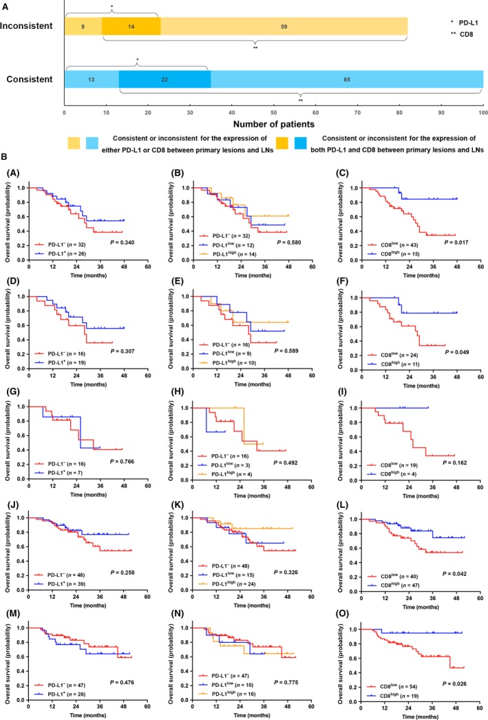
Figure 5.

(A) Bar chart showing the quantity of cases of consistent/inconsistent expression of PD‐L1/CD8+ TILs between PLs and LNs; (B) Kaplan–Meier survival curves of patients with LN+ (A–C), consistent expression of PD‐L1 (between PLs and LNs) (D–F), inconsistent expression of PD‐L1 (G–I), consistent expression of CD8+ TILs density (J–L), inconsistent expression of CD8+ TILs density (M–O). Other than patients with inconsistent expression of PD‐L1, CD8high TILs was associated with better OS than CD8low in the rest four groups. It showed no correlation between PD‐L1 expression and OS among all the five groups. Notes: LN+ refers to metastatic lymph nodes (patients staged as p‐TxN1‐3Mx). PLs, primary lesions; LNs, lymph nodes.
