
Figure 2.
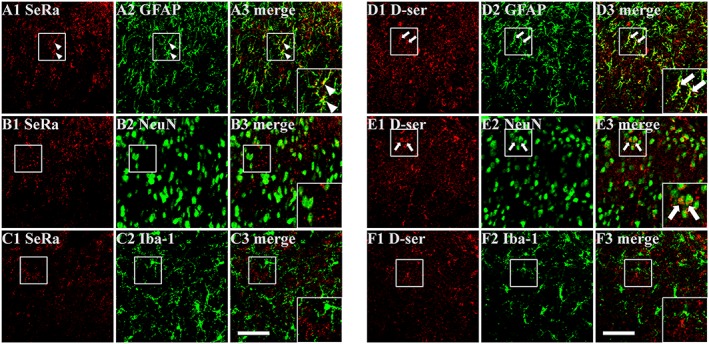
Representative photomicrographs depicting the cellular localization of serine racemase (SeRa) and D‐serine (D‐ser) immunoreactivity in the contralateral spinal cord dorsal horn at 10 days post‐carrageenan injection. (A–C) SeRa (A1, B1 and C1)‐immunoreactive cells were observed to co‐contain GFAP‐immunoreactivity (A3, arrow head), but not co‐contain NeuN (B3) or Iba‐1 (C3) immunoreactivity indicating that SeRa is localized to astrocytes. (D–F) D‐ser (D1, E1 and F1) immunoreactive cells co‐contained both GFAP (D3, arrow) and NeuN (E3, arrow) immunoreactivity indicating that both astrocytes and neurons contain D‐ser. Scale bar = 50 μm.