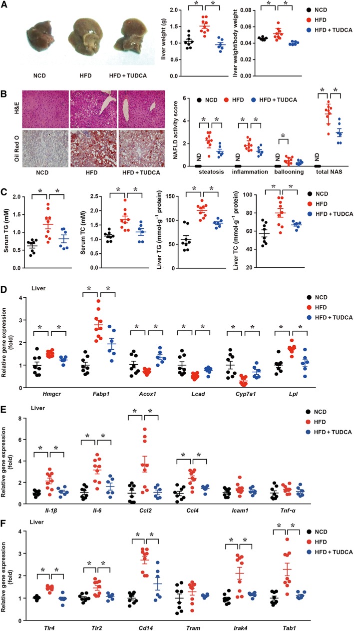
Figure 1.

TUDCA attenuates HFD‐induced hepatic steatosis and inflammatory responses in NAFLD mice. (A, left) Representative macroscopic images of the livers of NCD, HFD and TUDCA mice, (A, middle) Liver weight and (A, right) the ratio of the liver weight and body weight of the three groups. (B, left) Representative H&E‐treated and Oil Red O‐stained sections in liver samples and (B, right) NAS of three groups. (C) The serum and hepatic TG and TC contents of mice in the indicated groups were measured using the elisa. (D) The mRNA expression levels of genes associated with fatty acid synthesis, transport and β‐oxidation, (E) inflammatory cytokines and (F) innate immunity components in the liver were detected by qPCR. The data are presented as the means ± SEM. One‐way ANOVA followed by Newman–Keuls post hoc test for multiple comparison. NCD group, n = 8; HFD group, n = 9; and HFD + TUDCA, n = 6. *P < 0.05. Hmgcr, 3‐hydroxy‐3‐methylglutaryl CoA reductase. ND, not detected.
