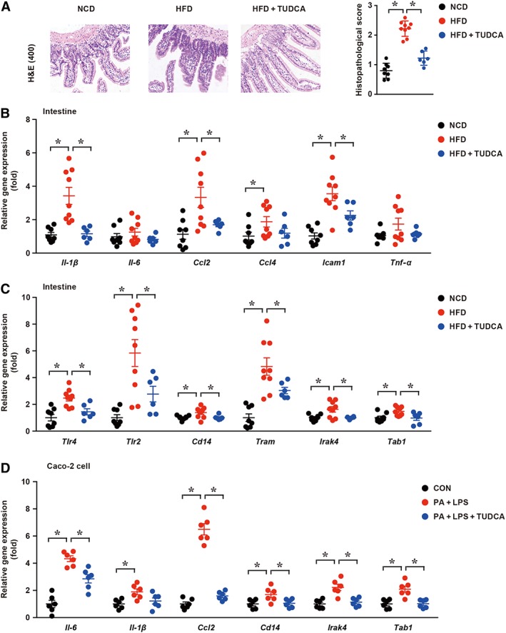
Figure 3.

TUDCA attenuates gut inflammation. (A) Representative ileum H&E staining sections (left panel) and corresponding histopathological score (right panel) in the NCD, HFD and HFD + TUDCA groups; (B, C) the mRNA expression levels of inflammatory cytokines and components of innate immune signalling were measured in the indicated groups; (D) the mRNA expression levels of inflammatory cytokines and components of innate immune signalling were measured in the indicated groups in Caco‐2 cells. CON, control group treated with vehicle; PA + LPS group, Caco‐2 cells co‐stimulated with 600 μM palmitate and 10 μg·mL−1 LPS for 6 h; PA + LPS + TUDCA group, Caco‐2 cells treated with the combination of palmitate (600 mΜ), LPS (10 μg·mL−1) and TUDCA (500 mM) for 6 h. The data are presented as the means ± SEM. One‐way ANOVA followed by the Newman–Keuls post hoc test for multiple comparisons. NCD group, n = 8; HFD group, n = 9; and HFD + TUDCA, n = 6. In vitro experiment, n = 5. *P < 0.05.
