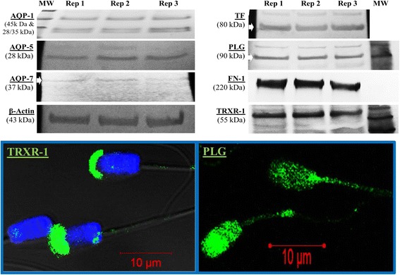
Fig. 2.

Representative westernblots and in situ immunofluorescence of boar spermatozoa. Selected proteins were targeted with antibodies raised against human AQP-1 (45 kDa & 28/35 kDa), AQP-5 (28 kDa), AQP-7 (37 kDa), β-actin (43 kDa, the internal loading control), TF (80 kDa), PLG (90 kDa), FN-1 (220 kDa), TRXR-1 (55 kDa). In situ immunofluorescence micrographs of TRXT-1 and PLG are also shown. The FITC green fluorescence indicates the sub-apical (acrosome membrane) localization of TRXR-1 on the sperm head and the localization of PLG on the head and mid-piece of spermatozoon. Nuclei are counterstained in blue with DAPI
