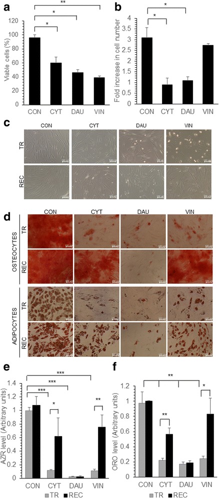
Fig. 1.

Effect of chemotherapeutic drugs on MSC proliferation and differentiation. MSC were either untreated (CON) or treated with CYT (10μm), DAU (0.1μm) and VIN (0.1μm) for 48 h. a The viable cells were counted microscopically using trypan blue exclusion and the percentage of viable cells was calculated. b Equal number of drug treated MSC were seeded in complete media without the drug, the viable cell number was counted after 7 days and the fold increase was calculated. c Representative microscopic images showing the morphology of MSC treated with the drugs for 48 h (TR) and drug treated cells allowed to proliferate in the drug free media for 7 days after drug treatment (REC). d Representative microscopic images showing the differentiation of MSC into osteoblasts and adipocytes either immediately after treatment with drugs (TR) or after the recovery of cells in drug free media for 7 days (REC). Osteogenic or adipogenic differentiation media was added to the cells to induce them to differentiate into respective lineage. e, f Alizarin red (AZR) or oil red O (ORO) quantification of MSC differentiated immediately after drug treatment (TR) or after recovery in drug free media (REC). AZR and ORO staining was done to detect osteogenic and adipogenic differentiation respectively. Values are mean ± SD, n = 3 samples. *p < 0.05, **p < 0.005, ***p < 0.0005
