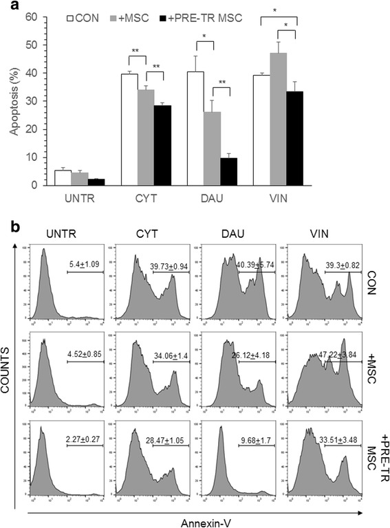
Fig. 3.

Chemoprotection of leukemia cells by drug treated MSC. a HL60 leukemia cells were cultured for 48 h in the absence of MSC (CON) or, in the presence of MSC (+MSC) or in the presence of drug pre-treated MSC (+PRE-TR MSC). The cells were treated with CYT (10μM), DAU (0.1μM) and VIN (0.1μM) for 48 h and apoptosis percentage was analyzed flow cytometrically by staining with annexin-V and PI. Values are mean ± SD, n = 3 samples. b Representative flow cytometric plots showing the apoptosis percentage in drug-treated HL60 as shown in (a). *p < 0.05, **p < 0.005
