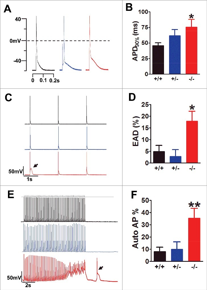
Figure 2.

Exon 33+/− cardiomyocyte did not show any signs of abnormal excitations. (A) AP waveforms recorded from WT (black), exon 33+/− (blue) and exon 33−/− (red) cardiomyocytes. (B) APD after 90% of repolarization (APD90%) from WT (n = 11 cells), exon 33+/− (n = 14 cells) and exon 33−/− (n = 16 cells) (*P<0.05 vs. WT, unpaired t test). (C and D) Detection of EADs in cardiomyocytes of WT (n = 73 cells, 17 mice), exon 33+/− (n = 24 cells, 5 mice) or exon 33−/− mice (n = 73 cells, 16 mice) at a 0.5-Hz pacing rate. (P = 0.0139 one-way ANOVA; *P<0.05 vs. WT, Bonferroni post hoc test). (E and F) Detection of autonomous APs in cardiomyocytes from WT (n = 73 cells, 17 mice), exon 33+/− (n = 24 cells, 5 mice) and exon 33−/− (n = 73 cells, 16 mice) a 5-Hz pacing rate (P = 0.0052, one-way ANOVA; **P<0.01 vs. WT, Bonferroni post hoc test).
