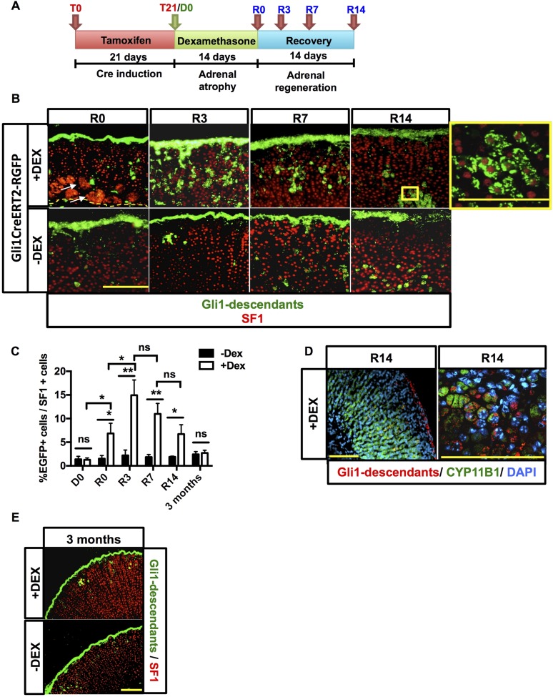
Figure 3.
Gli1 progeny participates in recovery of the adrenal gland. (A) Experimental scheme for treatment of Gli1CreERT2-R26REGFP mice. Following a tamoxifen chow diet for 21 days and administration of either dexamethasone or vehicle for 2 weeks, mice were euthanized at R0, R3, R7, and R14. (B) Immunofluorescence showing nuclear SF1 staining in red. Endogenous green EGFP marks Gli1+ cells and descendants. An increase in delamination of capsular Gli1+ cells into the adrenal cortex (green) that migrate centripetally toward the cortex-medulla boundary is present after dexamethasone (DEX) treatment. Capsular Gli1+ cells are SF1−, but their cortical progeny is SF1+, as shown in the enlargement box at the far top right. Scale bars: 100 μm. (C) Quantification of EGFP+ cells normalized to cortical Sf1+ cells in the cohorts of dexamethasone- and vehicle-treated mice. Error bars represent the standard error of the mean. *P < 0.05; **P < 0.01; and ***P < 0.001. (D) Gli1 descendant cells can differentiate into zF Cyp11b1+ cells. Scale bars: 100 μm. (E) Three-month chase of Gli1 descendants. For each time point, n = 5. Scale bars: 100 μm. ns, not significant.

