Figure 5.
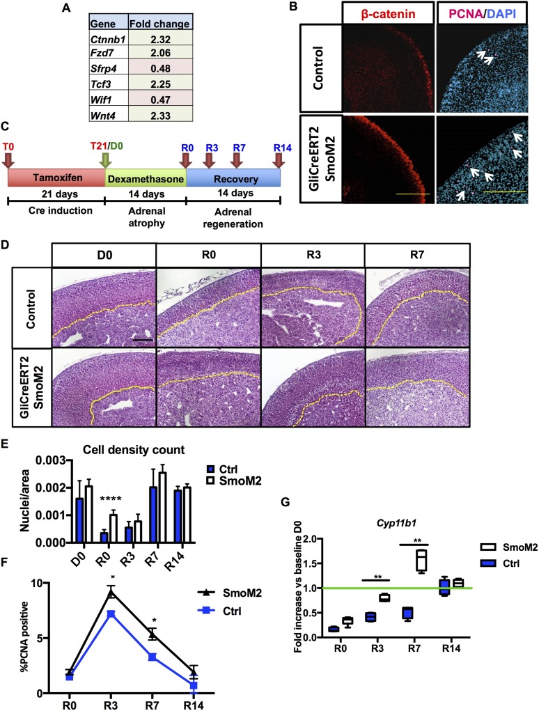
Constitutively active HH signaling in capsular Gli cells improves adrenal recovery. Analysis of Gli1CreERT2-SmoM2 mice: (A) qPCR array for canonical WNT pathway components detected differential expression of β-catenin (Ctnnb1), Fzd7, Sfrp4, Tcf3, Wif1, and Wnt4. (B) Immunofluorescence staining on 10-week-old male mice shows increased proliferation, as indicated by nuclear proliferating cell nuclear antigen (PCNA) in magenta (right panel, white arrows pointing at the PCNA-positive cells) and β-catenin expression in red (left panel). (C) Schematic of the experimental design. (D) Hematoxylin and eosin staining of control adrenals and adrenals expressing constitutively active smoothened (SmoM2 mice). Cortical medullary border marked in yellow. Scale bars: 100 μm. (E) Cell density count reveals higher cell density in SmoM2 cortices after dexamethasone treatment. (F) Quantification of proliferation shown as percentage of PCNA+ cells per field. (G) Relative quantification of Cyp11b1. For statistical analyses, all time points n = 5 animals, except R0, R3, R7, and R14 control groups (n = 6). Error bars represent the standard error of the mean. *P < 0.05; **P < 0.01; ***P < 0.001; and ****P < 0.0001.

