Figure 2.
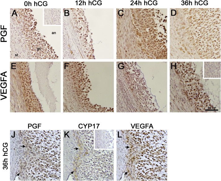
PGF and VEGFA immunodetection in monkey ovulatory follicles. Monkey ovaries obtained after ovarian stimulation in (A and E) the absence of hCG (0 hours) and (B and F) 12, (C and G) 24, or (D and H) 36 hours after hCG were used for immunodetection of (A–D) PGF or (E–H) VEGFA as indicated by presence of brown precipitate. Additional serial sections from an ovary obtained after ovarian stimulation and 36 hours hCG were used for immunodetection of (J) PGF, (K) the theca cell enzyme CYP17, and (L) VEGFA to confirm colocalization of PGF and VEGFA with CYP17 (arrows). Tissue sections were counterstained with hematoxylin (blue). Insets show absence of brown stain was confirmed when primary antibodies were omitted for (A) PGF, (H) VEGFA, and (K) CYP17. All images are at same magnification and use bar in panel H (50 µm). All images are oriented as in panel A, with stroma (st) in lower left, granulosa cells (gc) central, and antrum (an) in upper right. Images are representative of n = 3 to 4 monkeys per group.

