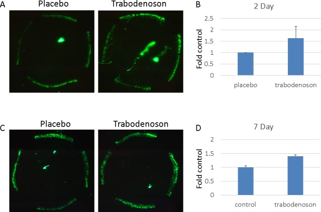
Figure 4.
Trabodenoson effects on tracer deposition in conventional outflow tissues. The experiments were conducted in two groups of young mice. One group of mice was treated daily with trabodenoson topically on one eye and placebo on the contralateral eye for two consecutive days. The second group was treated topically daily for 7 days. At end of the study, the mice were anesthetized and a fixed amount of fluorescent beads diluted in DBG were perfused simultaneously into paired eyes at a constant flow rate. Fluorescence intensity of tracer deposited in outflow tissues was visualized and captured on flat-mounted anterior segments using an epifluorescence microscope. (A, C) Representative flat-mounts from eyes treated for 2 and 7 days, respectively. (B, D) Combined data (mean ± SE). n = 8 for 2-day treatment group (B) and n = 6 for 7-day treatment group (D).

