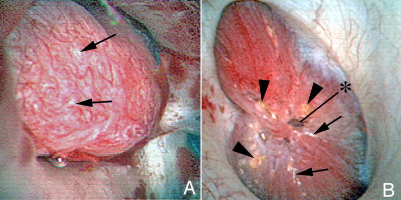
Figure 1.
A: A papillae from an ICSF patient showing a small amount of Randall’s plaque (arrows). The papillae is otherwise normal appearing.
B: A papillae from a BRSF patient. The papillae is severely diseased. It is flattened. In the center of the papillae is a grossly dilated Bellini duct (asterisk). In addition to Randall’s plaque (arrows), Bellini duct deposits are seen (arrowheads).

