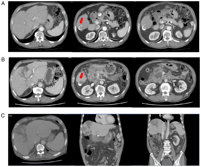
Figure 1.
(A) Liver dynamic CT performed 2 days before the TACE with DC beads showing a viable remnant tumour (segment 6, arrow). (B) Liver dynamic CT performed on admission due to abdominal pain revealed multi-locular fluid accumulation with enhanced walls in the peripancreatic space and marked irregular right intrahepatic duct dilatation. However, imaging indicated increased necrosis (3 cm) in the lipiodol-laden hepatocellular carcinoma at segment 6. (C) Liver dynamic CT re-performed for fever and abdominal pain after initial improvement (44 days after the TACE with DC beads) revealing newly developed multifocal perfusion defects, suggesting liver and splenic infarction. The peri-pancreatic fluid collection was decreased. DC beads, drug-eluting beads; CT, computed tomography.

