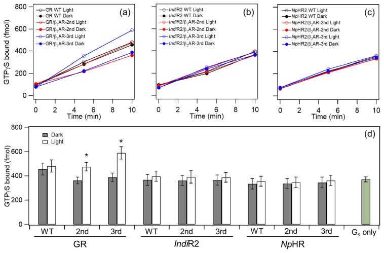
Figure 3.
G-protein activation by GR chimeras (a), IndiR2 chimeras (b), and NpHR chimeras (c). Time-dependent GTPγS-binding to Gs-protein was monitored in the light (open circle) and dark (filled circle). Black, red and blue circles/lines represent the results of WT, the second and third loop chimera of β2AR, respectively. (d) Comparison of G-protein activation ability by WT and chimeras. GTPγS-binding to Gs-protein was monitored at 10 min in the light (open bar) and dark (filled bar). It should be noted that the spontaneous incorporation of GTPγS of Gs (Gs only) was about 40 times higher than that of Gt [17,18]. Data are presented as the means±S.D. of more than three independent experiments and the marked chimeras (*) exhibit a significant difference between light-dependent and dark activations (p<0.05; Student’s t-test, one-tailed).

