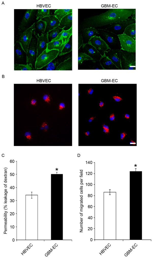
Figure 1.
Increased permeability and motility in GBM-EC. (A) Cluster of differentiation 31 (green) immunostaining of primary isolated brain ECs from normal HBVECs and GBM-ECs. Nuclei were stained with DAPI (blue). (B) Accumulation of acetylated low-density lipoprotein (red) by primary HBVECs and GBM-ECs. (C) HBVECs and GBM-ECs were grown to confluence on Transwell filters. Permeability for FITC-dextran (70 kDa) was measured using a fluorescence plate reader (λ excitation 485 nm; λ emission 530 nm). (D) A Transwell motility assay using HBVCEs and GBM-ECs was performed using 8-µm Transwell chambers. Migrated cells were quantified by counting crystal violet stained cells in 4 random fields of view/filter. Results are the mean ± standard error of three independent experiments. Scale bars, 10 µm. *P<0.05 vs. HBVECs. HBVEC, human brain vascular endothelial cells; GBM-ECs, glioblastoma endothelial cells; FITC, fluorescein isothiocyanate.

