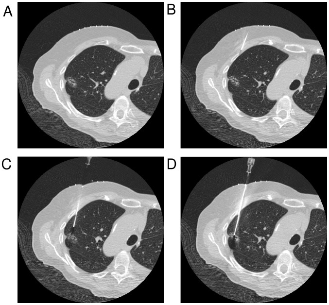
Figure 2.
Conventional CT-guided biopsy method. (A) The center of the lesion was positioned at the CT landmark using a radiopaque grid placed on the patient's skin. (B) The initial puncture was stopped when the surface of the pleura was reached. Biopsies were performed to avoid ribs, bullae, vessels, and fissures. (C) If the lesion was on the extended course of the needle track, the biopsy procedure was continued. If the nodule was not on the extended course of the needle track, the course or puncture site was changed. (D) Specimens obtained were immediately immersed in 10% buffered formalin solution.

