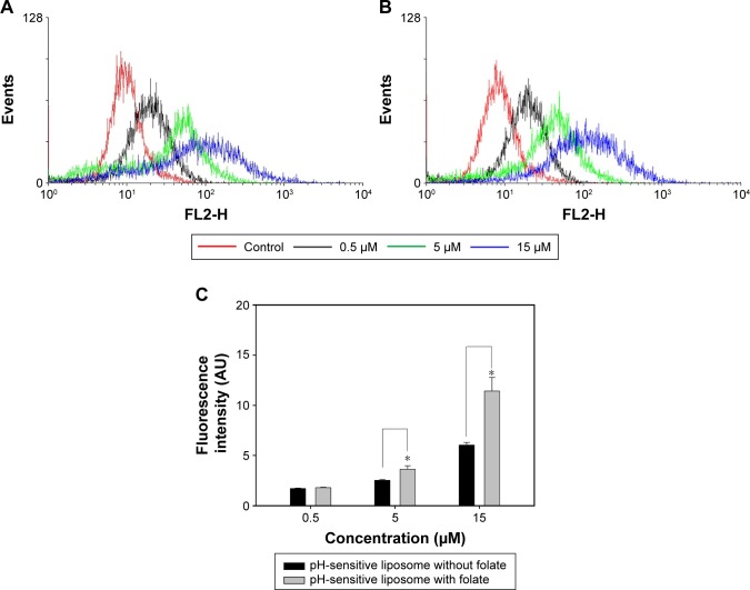
Figure 5.
For flow cytometric analyses, MCF-7 cells were treated for 24 h with 0.5, 5, and 15 µM of pH-sensitive liposomes (A) with or (B) without a folate modification. (C) Fluorescence intensities were quantified as cellular uptake. Control was a cell blank without any treatment.
Notes: Data are presented as mean ± standard deviation (n=3). *p<0.05 was considered statistically significant.

