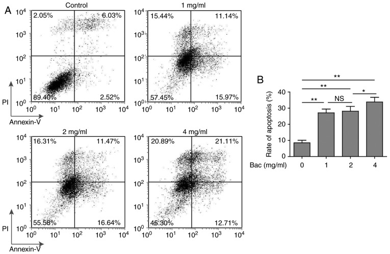
Figure 2.
The pro-apoptosis effect of bevacizumab on glioblastoma cells. U87-MG cells were treated with different concentrations of bevacizumab for 48 h, and then the cell apoptosis was assessed by Annexin V/PI staining and flow cytometry analysis. (A) One representative FACS result from three independent experiments. (B) The rate of apoptosis for cells in A. Error bars represented mean ± SD. P-values were determined by one-way ANOVA followed by Tukeys post hoc test. **P<0.01, *P<0.05 and ns, not significant. ANOVA, analysis of variance.

