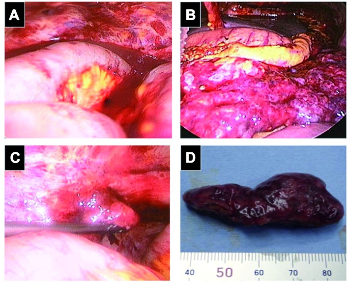
Figure 2.
Laparoscopic findings. (A) Massive bloody ascites and peritoneal inflammation were observed. (B) A congested omental mass invaded close to the splenic hilum. (C) A small papillary lesion (arrow) is seen on the surface of the left Fallopian tube. (D) Macroscopic findings of the biopsied omental mass.

