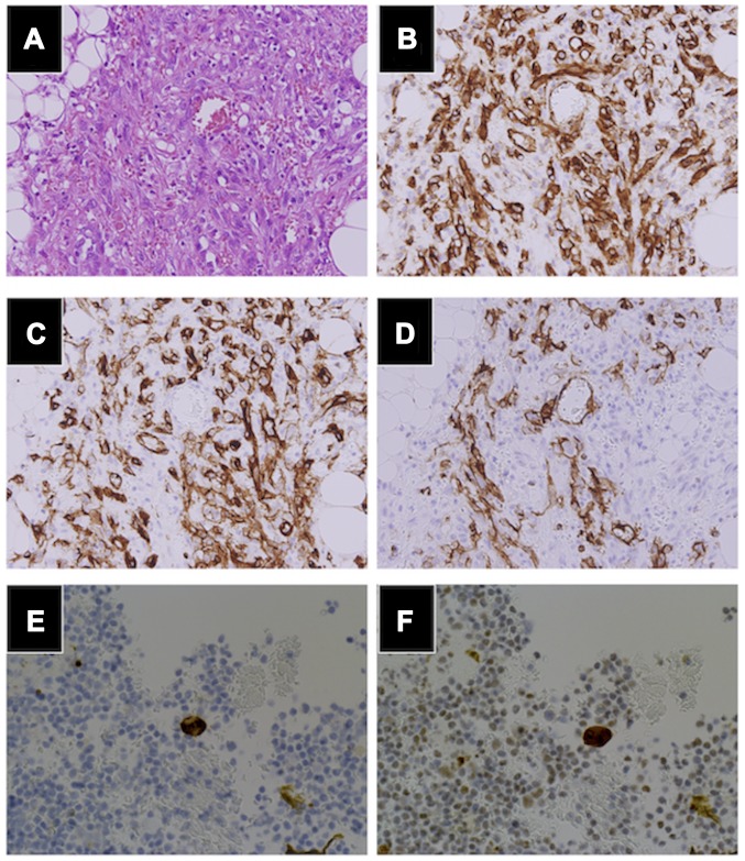
Figure 3.
Histopathological findings. (A) H&E stains of the omental mass showed high-grade malignant cells with frequent mitosis, forming irregular anastomosing vascular channels. Immunohistochemistry was positive for (B) CD31, (C) CD34 and partially positive for (D) D2-40, while pan-keratin was grossly negative. The cell block method was used for immunohistochemical investigation of ascites, and showed a small number of cells positive for (E) CD34 and (F) ERG. (A-C) Magnification, ×20; (E-F) Magnification, ×40.

