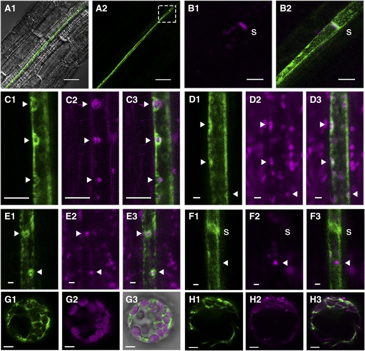
Figure 5.
Localization of SLI1 in Arabidopsis.
(A1) to (B2) Confocal images of pSLI1:EYFP:SLI1 (green) in seedling roots ([A1] and [A2]), with details in the boxed region showing aniline blue staining (magenta) of a sieve plate (B1) and merged signals of aniline blue and SLI1 (B2).
(C) FM4-64 staining (30 h incubation; 1, SLI1; 2, FM4-64; 3, merged).
(D) MitoTracker staining (1, SLI1; 2, MitoTracker Deep Red; 3, merged).
(E) Cotransformation with the mitochondria marker ScCOX4 (Nelson et al., 2007) (1, SLI1; 2, ScCOX4:mCherry; 3, merged).
(F) Cotransformation with the plastid marker tpCab (Kim et al., 2013) (1, SLI1; 2, tpCab:mCherry; 3, merged).
(G) Arabidopsis protoplast with transient expression of 35S:EYFP:SLI1 (1, SLI1; 2, chloroplasts; 3, merged).
(H) Protoplast stained with ER Tracker Red (1, SLI1; 2, ER Tracker; 3, merged).
Images represent consistent results in at least two independent transgenic lines. s, sieve plate; solid arrows, phloem organelle location. Bars = 25 µm in (A), 5 µm in (B) and (C), 1 µm in (D) to (F), 5 µm in (G) and (H).

