Figure 3.
Inhibition of Exocyst Function Results in Growth Inhibition and PIN2-GFP Trafficking Defects in scd1 and scd2 Mutants.
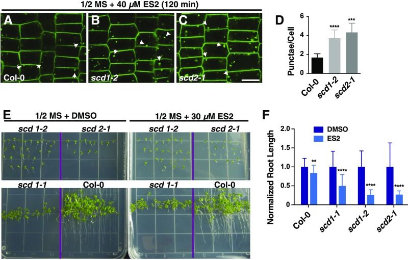
(A) to (C) PIN2-GFP in wild-type (A), scd1-2 (B), and scd2-1 (C) roots from 5-d-old seedlings treated for 2 h in 0.5× MS plus 40 µM ES2. Arrowheads indicate PIN2-GFP-labeled intracellular accumulations. Bar = 10 µm.
(D) Quantitation of average PIN2-GFP-labeled ES2-induced punctae per cell. Shown are means ± sd. ***P < 0.001 and ****P < 0.0001 (t test, compared with wild-type value).
(E) Seedlings grown on agar plates with 0.5× MS or 0.5× MS plus 30 µM ES2.
(F) Quantitation of relative root length of wild-type and scd1-2 and scd2-1 mutants grown on 0.5× MS agar plates containing DMSO or 30 µM ES2. Shown are means ± sd. **P < 0.01 and ****P < 0.0001 (t test, compared with DMSO value).

