Figure 2.
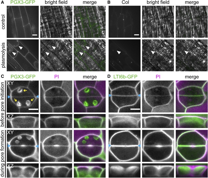
In Seedlings, PGX3-GFP Is Localized in the Cell Wall and Accumulates at Stomatal Pore Initiation Sites.
(A) and (B) Fluorescence and bright-field images of 5-d-old roots. Images show roots expressing ProPGX3:PGX3-GFP (A) or Col roots (B) under the control condition (top panel) or plasmolyzed with 1 M mannitol for 5 min (bottom panel). White arrowheads indicate membranes separated from the cell wall.
(C) and (D) PI staining in developing guard cells of 4-d-old seedlings. Images show seedlings expressing ProPGX3:PGX3-GFP (C) or a membrane marker, GFP-tagged LTI6b (D). Blue arrowheads indicate cell plates between sister guard cells. Yellow arrows indicate autofluorescence from chloroplasts. XY and XZ indicate projections in the XY and XZ directions, respectively.
Bars = 10 µm in (A) and (B) and 5 µm in (C) and (D).

