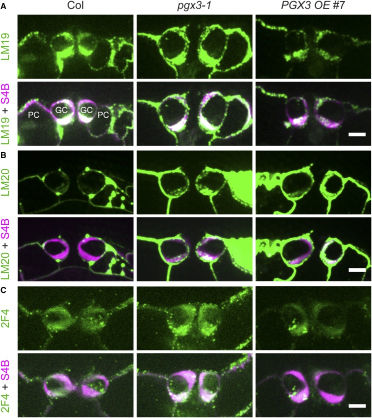
Figure 7.
Immunolabeling in Guard Cell Walls Reveals Changes in Antibody Binding.
(A) Colabeling of LM19, an antibody that recognizes demethylesterified HG, and S4B, a dye that binds to cellulose, in cross sections of guard cell pairs in 3- to 4-week-old Col, pgx3-1, and PGX3 OE #7 plants. Top panels, LM19 labeling; bottom panels, LM19 labeling (green) merged with S4B signals (magenta) in the same guard cell pair. GC, guard cells; PC, pavement cells.
(B) Colabeling of LM20, an antibody that recognizes methylesterified HG, and S4B in cross sections of guard cell pairs in 3- to 4-week-old Col, pgx3-1, and PGX3 OE #7 plants. Top panels, LM20 labeling; bottom panels, LM20 labeling (green) merged with S4B signals (magenta) in the same guard cell pair.
(C) Colabeling of 2F4, an antibody that recognizes Ca2+-cross-linked HG, and S4B in cross sections of guard cell pairs in 3- to 4-week-old Col, pgx3-1, and PGX3 OE #7 plants. Top panels, 2F4 labeling; bottom panels, 2F4 labeling (green) merged with S4B signals (magenta) in the same guard cell pair.
Bars = 5 µm in (A) to (C).

