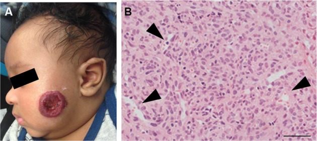
Figure 1.

(A) A proliferating IH on the cheek of an infant. Note the ulceration in the center of the IH. (B) A proliferating IH is highly cellular with poorly defined vascular spaces (black arrowheads).
Note: Magnification of image B, 40×; scale bar =50 µm.
Abbreviation: IH, infantile hemangioma.
