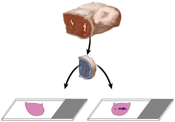
Fig. 4.
Unequivocal orientation of the revised tumor bed margin. The new margin surface of the revised anterior margin from left partial glossectomy is indicated by ink. The white irregular area in the anterior aspect of glossectomy specimen represents residual carcinoma at the initial anterior margin. The dark blue irregular area on the right glass slide represents tumor. Without orientation, the surface to be first examined intraoperatively is picked randomly. When indicated, the new margin surface will be examined first intraoperatively. If the frozen section is negative for tumor, but the permanent section of the frozen remnant reveals tumor, the overall margin status is “close, but negative”. Such determination is impossible if the tumor bed margin was fragmented or the new true margin surface was not indicated. In addition to ink, new margin surface can be indicated by a stitch, clip, or directly by surgeon. This revision margin is thin and is best processed as shave margin. Thicker margins can be processed as radial margins (new margin surface re-inked, cut into, and embedded on edge).

