
Fig. 5. MEIS1/HOXA9 binds to a predicted consensus site in the enhancer E9.
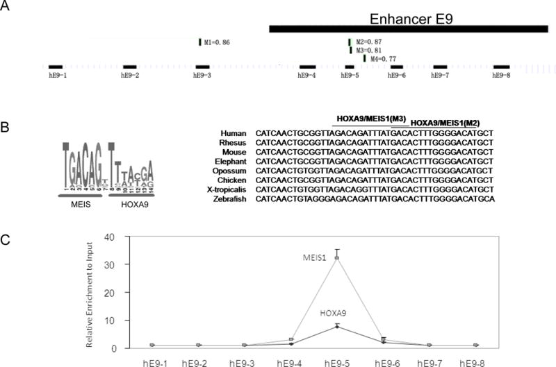
A). Schematic map of the enhancer E9, predicted MEIS1/HOXA9 binding sites and the sequence alignment of the detected PCR replicon in ChIP assay. Four HOXA9/MEIS1binding sites (M1 to M4) were predicted by bioinformatics analysis (http://www.genomatix.de) with the simultaneous matrix shown (Also see Supplementary Table S2).
B). HOXA9/MEIS1 binding motif by Genomatix and the conserved sequence of MEIS1/HOXA9 binding sites from various vertebrate species.
C). Crosslinked chromatin from THP-1 was immunoprecipitated with antibodies to MEIS1 and HOXA9. The precipitated DNA was amplified using the primer pairs spanning the enhancer E9 region. A higher binding signal was detected with the primer pair hE9-5.