
Fig. 4. G1T28-induced PQ protects HSCs from proliferative exhaustion.
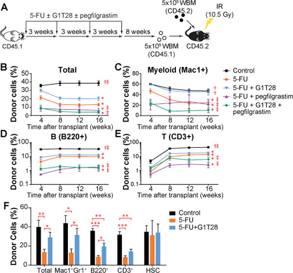
(A) Treatment and BM transplantation schematic. WBM, whole bone marrow cells (B-E) The percentage of donor-derived cells (CD45.1+) in total PB leukocytes (B), or myeloid cell (Mac1+, C), B lymphocyte (B220+, D), and T lymphocyte (CD3+, E) fractions 1 to 4 months after primary BM transplantation. 3-8 donor mice/treatment, 5 recipients/donor. Statistical significance of differences between treatment groups at 16 weeks after transplantation was assessed using one-way ANOVA with correction for multiple comparisons. (*, P < 0.05 compared to control; †, P < 0.05 compared to 5FU + vehicle, ‡, P < 0.05 compared to 5FU + G1T28). (F) The percentages of donor-derived cells in total BM cells, HSCs, and major BM cell lineages 32 weeks after BM transplantation. 5 donor mice/treatment, 5 recipients/donor. Statistical significance was assessed by two-tailed Student’s t-test (*P < 0.05, **P < 0.01, ***P < 0.001). Average donor cell reconstitution in B-F was calculated by first averaging the value of the 5 recipients from each donor, followed by calculating the mean of all donors. Error bars represent SEM.