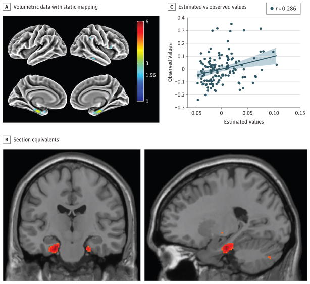
Figure 2. Association of Subjective Cognitive Decline (SCD) With Flortaucipir F 18 Positron Emission Tomography (FTP-PET) Findings.
Whole-brain voxel wise analysis with FTP-PET (previously known as AV 1451, T807) standardized uptake value ratio (SUVR) was performed after accounting for age. A, Surface rendering of volumetric data with static mapping from Montreal Neurological Institute (MNI) space to fs average from FreeSurfer software. B, Brain section equivalents are shown to better visualize the medial temporal lobe (chosen from apex t value in the region of interest; ie, MNI coordinates −22 − 18 − 26). Color bar indicates t values. C, Plot of the estimated and observed values from the apex of the entorhinal cortex region of interest from the general linear model of FTP-PET SUVR approximated as SCD + age. Diagonal lines indicate lines of best fit for the correlational analysis; shading, 95% CI; and data points, study participants.

