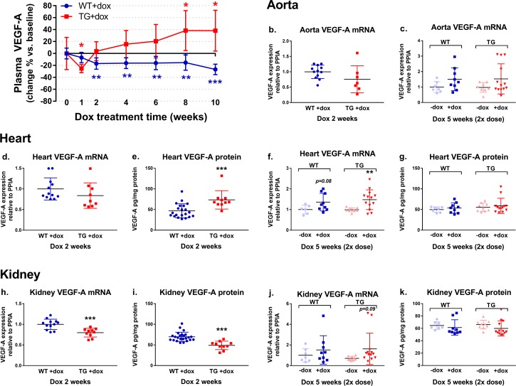
Fig 3.
Plasma VEGF-A concentration in response to dox treatment (a). Tissue VEGF-A mRNA and protein levels after two weeks of 1 mg/ml dox treatment and five weeks of 2 mg/ml dox dose treatment (b-k). Dox treatment decreased plasma VEGF-A levels in TG mice after 1 week, after which the plasma VEGF-A increased. In WT mice a decreasing effect on plasma VEGF-A was seen (a). *p<0.05, **p<0.01 and ***p<0.001 compared to baseline within each group, 1-way ANOVA with Dunnett´s post hoc test, n = 9-10/group. In the selected tissues, aorta, heart and kidney, the dox treatment with the 1 mg/ml dox dose for 2 weeks showed decreasing trend in VEGF-A mRNA expression in TG mice in comparison to WT mice (b, d, h), which was associated with increased cardiac VEGF-A protein (e) and decreased kidney VEGF-A protein levels (i). When the dox dose was doubled and the treatment time increased to 5 weeks, a trend towards increasing VEGF-A expression was seen in all three tissues in both WT and TG mice (c, f, j). However, no changes were detected in protein levels (g, k). **p<0.01 and ***p<0.001 compared to WT+dox group in 2 weeks experiment (b, d, e, h, i) or to no dox group (-dox) in 5 weeks experiment (c, f, g, j, k), t-test, n = 6-24/group.

