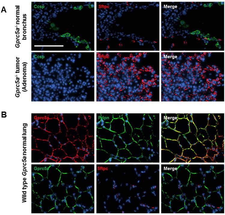
Figure 3. Immunofluorescence analysis of airway lineage markers in lung lesions and normal tissues.

Immunofluorescence (IF) analysis of airway lineage markers of Clara (Ccsp) and alveolar type 2 (AT2) (Sftpc) cells was performed in Gprc5a-/- lesions and adjacent normal regions as described in Materials and Methods. B. IF analysis of Gprc5a and the AT1 cell marker Pdpn was performed as described in Materials and Methods. All images were captured using a BX61 immunofluorescent system (Olympus) and merged using the CytoVision workstation (Leica biosystems Inc.) at a magnification of 200× (scale bar denoting 100 μm).
