
FIG 2.
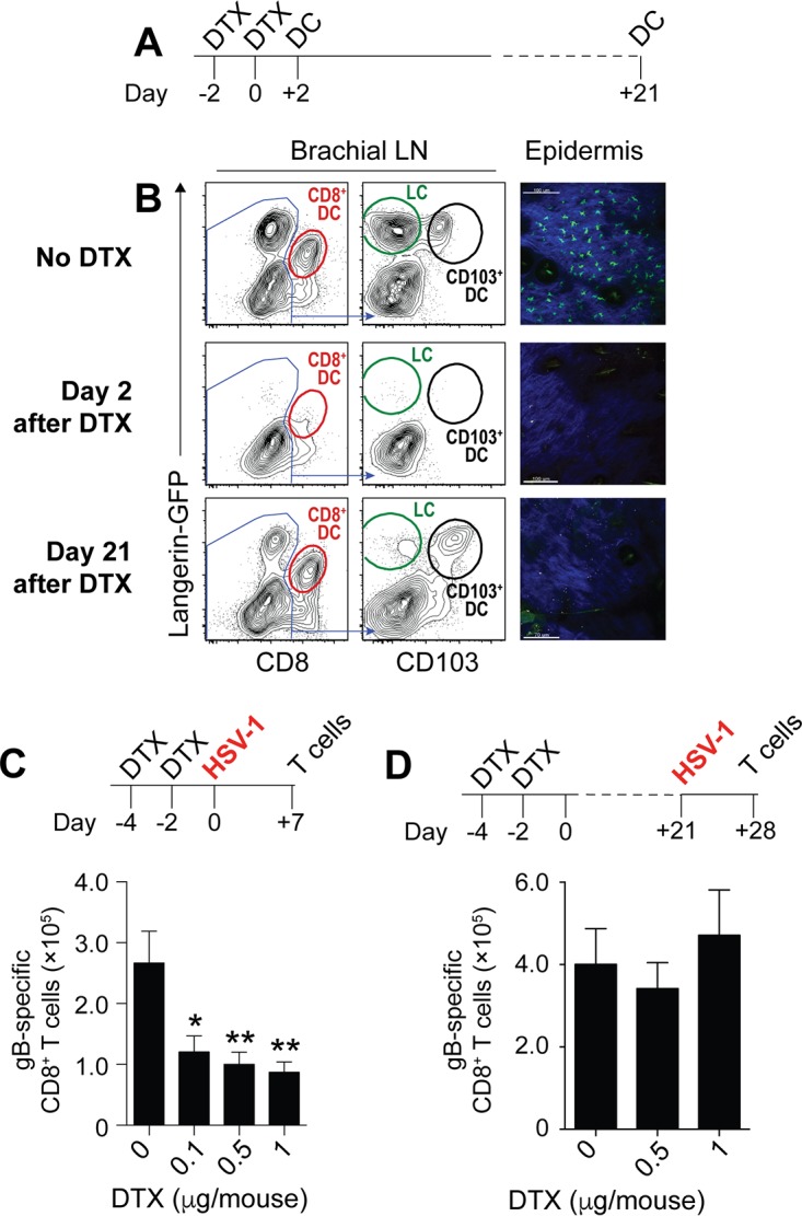
Langerin-positive DC, but not LC, are required for HSV-specific CD8+ T cell priming. (A) Schematic depicting the DTX treatment regimen for the assessment of DC depletion. Mice were treated with DTX on days −4 and −2. DC in the brachial LN and the epidermis were analyzed 2 days or 21 days after the last DTX treatment. (B) Representative plots of MHC-IIhi CD11chi cells enriched from the brachial LN on day 2 or day 21 after DTX treatment on two consecutive days. Cells were first plotted based on expression of CD8 and GFP (langerin) to identify CD8+ DC. The CD8− fraction was then further divided into LC (GFP+ CD103−) and CD103+ DC (GFP+ CD103+). (C and D) Top panels, schematics depicting the DTX treatment regimen to determine its impact on HSV-specific CD8+ T cell priming. Mice were treated with DTX on days −4 and −2. On day 0 (C) or day 21 (D) mice were infected on the skin with HSV-1, and the spleens were analyzed for virus-specific CD8 T cells 7 days later. Bottom panels, absolute numbers of gB498–505-specific CD8+ T cells in the spleen on day 7 after HSV-1 skin infection of Lg-DTR mice that last received DTX 2 days (C) or 21 days (D) earlier relative to those in PBS controls. All data are pooled results from at least two independent experiments (n = 3 to 5 per experiment) and are expressed as mean + SEM. Asterisks indicate statistically significant differences versus controls as assessed by one-way analysis of variance (ANOVA) (*, P < 0.05).