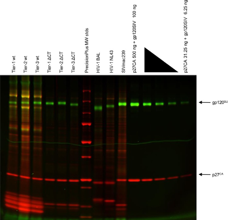
FIG 3.
Quantification of virion Env content using dual-fluorescence imaging of virus proteins. A66-R5-derived test viruses, SupT1-R5-derived control viruses, and a series of protein standards were run on an SDS-PAGE gel. The gel was stained with the Pro-Q Emerald 300 glycoprotein dye to detect gp120 and with the SYPRO ruby dye to detect total protein, including p27. MW, molecular weight.

