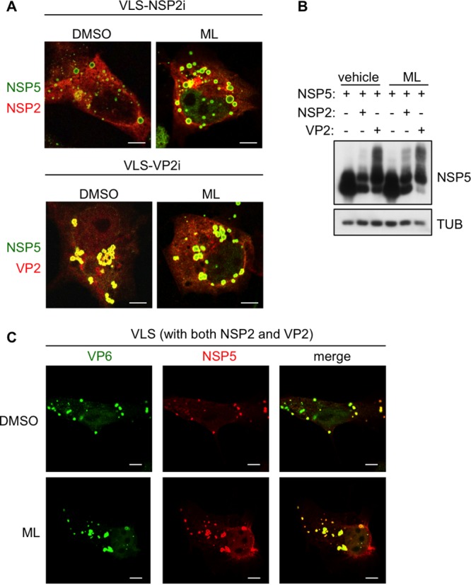
FIG 7.

ML effect on VLS. Confocal immunofluorescence assay of VLS (A and C) and Western blot analysis (B) with the indicated antibodies of MA104 cells transfected with NSP5, NSP2, VP2, and VP6, as indicated. (A) NSP5 is shown in green and NSP2 or VP2 in red. (C) NSP5 is shown in red and VP6 in green. Cells were treated for 5 h with 10 μM ML or DMSO at 18 h posttransfection. Bars, 5 μm.
