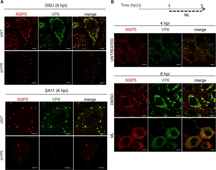
FIG 8.
VP6 in RV-infected cells. Shown is confocal immunofluorescence of MA104 cells infected with either OSU or SA11 (MOI, 25 VFU/cell) and transfected with siRNAs specific for SA11 VP6 or OSU VP6 or with a nontargeting siRNA (siNT) (A) or treated with 10 μM ML or DMSO (B). At the indicated times postinfection, viroplasms were visualized with anti-NSP5 antibody (red) and VP6 with MAb 4B2D2 (green). Scale bars, 5 μm.

