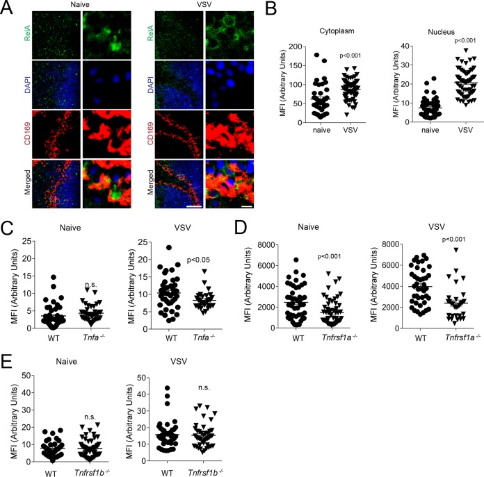
FIG 6.
VSV infection leads to TNFR1-dependent canonical NF-κB activation in splenic CD169+ cells. (A to D) Sections of snap-frozen spleen tissue were harvested 4 h after infection with 2 × 10⁸ PFU VSV. (A) The sections were stained for RelA before and after infection (one representative result out of 3 is shown; scale bar = 100 μm). Enlarged images of the boxed areas in the merged images on the left side are shown on the right (scale bar = 5 μm). (B) MFI of cytoplasmic and respective nuclear RelA quantified in CD169+ cells from WT mice before and after VSV infection to evaluate nuclear translocation of RelA (n = 48 to 63). (C to E) Spleen sections from WT and Tnfa−/− (C), Tnfrsf1a−/− (D), and Tnfrsf1b−/− (E) mice were stained with anti-RelA antibodies 4 h after infection with 2 × 10⁸ PFU VSV. The MFI of RelA in the nuclei of CD169+ cells was determined with ImageJ software (n = 35 to 57). The error bars indicate SEM.

