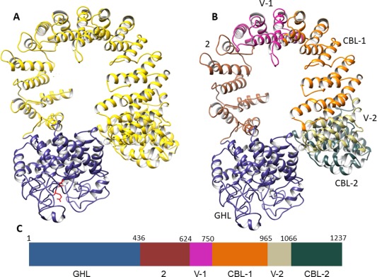
Figure 1.

Theoretical 3‐D structure of the PhK α subunit. (A) Hierarchical protein structural modeling of the α subunit carried out using I‐TASSER.20 X‐ray crystal structures of glucodextranase (PDB ID: 1ULV) from Anthrobacter globiformis bound to acarbose (not shown) and importin β (IBL)(PDB ID: 4C0O) from human were used to thread, respectively, residues 1–436 (blue‐gray ribbon trace) and 437–1237 (gold trace) of the multi‐domain α sequence. Putative catalytic Glu residues 185 and 371 are shown in red. (B) Subdomains of the large IBL domain labeled and color coded to match the schematic of the linear structure of α shown in (C). Further modeling details of the GHL domain and IBL subdomains are listed in Table I.
