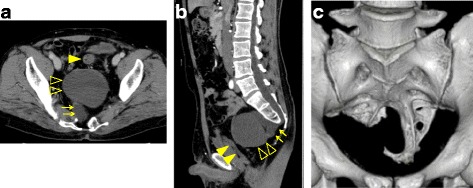
Fig. 1.

Computed tomography (CT) scan showing a 60 × 30 × 21-mm meningocele (→) which was protuberated anteriorly from the level of S3; a 90 × 85 × 68-mm mass (△) which was located anterior to the meningocele; and a dislocated rectum (▲) (a, b). A partial defect of the sacrum with deformity was detected (c)
