
Fig. 4.
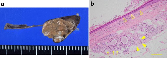
Macroscopic appearance (a) and microscopic findings (b) of a section of the resected teratoma. The cyst wall was mainly composed of keratinizing stratified squamous epithelium (△). Fat, sweat glands (arrows), and peripheral nerves (▲) were observed around the cyst wall, and the pathological diagnosis was mature teratoma. The scale bar represents 200 μm