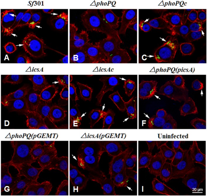
Figure 2.
Changes in the cytoskeleton of HeLa cells infected with S. flexneri strains. HeLa cells were infected with S. flexneri strains for 15 min. Actin was visualized by staining with Texas Red-labeled phalloidin (red), bacteria were stained with rabbit polyclonal anti-Shigella anti-serum (green), and nuclei of HeLa cells and bacterial DNA were stained with DAPI (blue). The coverslips were mounted and observed under a confocal laser scanning microscope at ×100 magnification (A–I). Arrows indicate locations of membrane ruffles. The ΔphoPQ(pGEMT) and ΔicsA(pGEMT) were used as empty plasmid controls.

