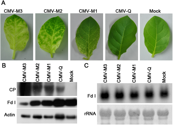
Figure 3.
Detection of Fd I levels following an infection by CMV. (A) Leaves exhibiting severe chlorosis symptoms, mild chlorosis symptoms, or vein-clearing symptoms from CMV-M–infected Nicotiana tabacum cv. Samsun plants, and asymptomatic leaves from tobacco plants infected by CMV-Q and mock-inoculated plants. (B) Western blots using anti-CP monoclonal and anti-Fd I polyclonal antibodies to analyse CMV CP and Fd I in the abovementioned samples, with actin as a control. (C) Northern blot conducted with 10 µg total RNA to examine the accumulation of Fd I transcription using a specific probe corresponding to position 75–435 of Fd I mRNA. The loading control comprised 25 S rRNA.

