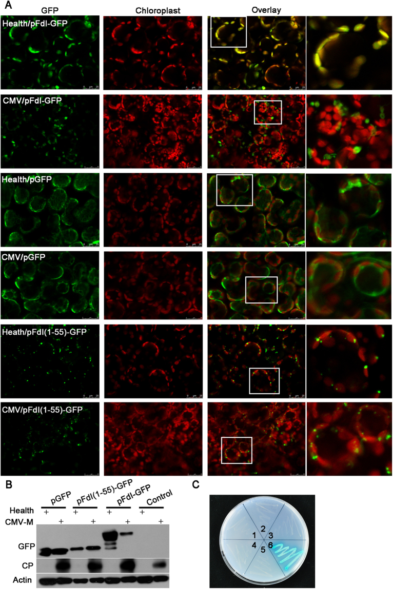
Figure 5.
Subcellular production and localization of Fd I in CMV-M–infected or healthy Nicotiana benthamiana plants. (A) The Fd I-GFP fusion protein mainly localized in the chloroplasts and cytoplasm of healthy and CMV-M–infected plants, respectively. Whereas, GFP alone was mainly localized in the cytoplasm in healthy and CMV-M–infected hosts. Residues 1–55 at the N-terminal of Fd I formed a transit peptide targeting GFP into chloroplasts in both healthy and CMV-M–infected plants. Scale bars = 25 µm. (B) Western blot analysis with an anti-GFP antibody to detect GFP alone (pGFP), GFP fused to Fd I (pFdI-GFP), and GFP fused to the Fd I transit peptide [pFdI(1–55)-GFP]. (C) In yeast cells, the CP of CMV-M did not interact with the Fd I transit peptide or mature Fd I. 1, pGADT7-RecT/pGBKT7-Lam (negative control); 2, pGAD-FdI (1–55)/pGBKT7; 3, pGAD-FdI (56–145)/pGBKT7; 4, pGAD-FdI (1–55)/pGBK-MCP; 5, pGAD-FdI (56–145)/pGBK-MCP; 6, pGADT7-RecT/pGBKT7-53 (positive control).

