Figure 4.
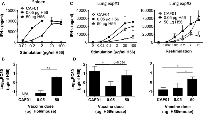
High dose post-exposure H56 vaccination leads to reduced functional avidity. One week post the final vaccination in the post-exposure model, splenocytes (A) or lung cells (C) from control (open circle) and H56-vaccinated (0.05 µg closed square; 50 µg open triangle) mice were cultured for 3 days with varying concentrations of H56, and IFN-γ release in the culture supernatants was measured by ELISA. (A) Data points represent mean ± SD of triplicate cultures of pooled cells from three mice/group (from Exp. 4 in Figure S1B in Supplementary Material). (B) EC50 was calculated using data from (A); bars, mean ± SD per vaccine group. This experiment was repeated twice. (C,D) IFN-γ release and calculated functional avidity (EC50) of lung cells from two independent experiments in which subsequent week 37 protection was not assessed. (C) Data points represent mean ± SD of triplicate cultures pooled from five mice/group. (D) Bars indicate mean ± SD EC50 calculated from (C). Statistical differences between functional avidity of vaccine groups were assessed by a one-way ANOVA and Tukey’s posttest for multiple comparisons. *p < 0.05, **p < 0.01. No further identical experiments with lung T cell avidity were performed.

