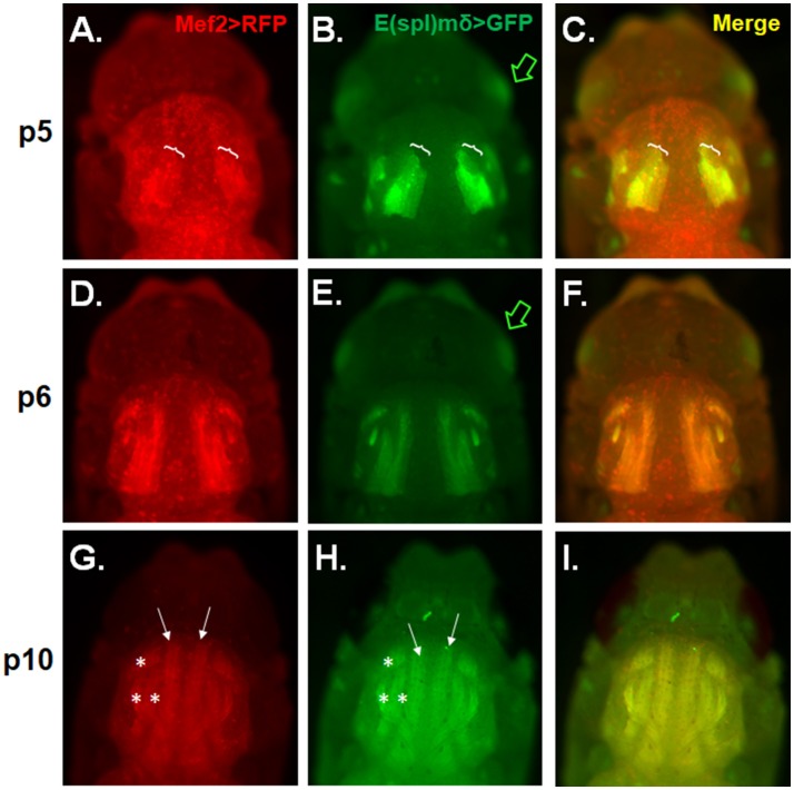
Figure 4.
E(spl)mδ is expressed in the developing IFM. Live epifluorescent imaging of developing IFMs in pupae carrying both Mef2-RFP and E(spl)mδ-GFP at the indicated stages (A–C) p5, (D–F) p6, and (G–I) p10. (C,F,I) Merged images of RFP and GFP represent overlapping regions of expression of Mef2 in developing IFMs and E(spl)mδ. Pupae were dissected from their pupal cases and imaged directly. Asterisks mark the dorsal ventral muscles (DVM) and closed arrows mark the dorsal longitudinal muscles (DLM). Brackets mark the regions of DLM development that encompass myoblast fusing to the larval templates. Green arrow points to E(spl)mδ expression in the developing eye.

