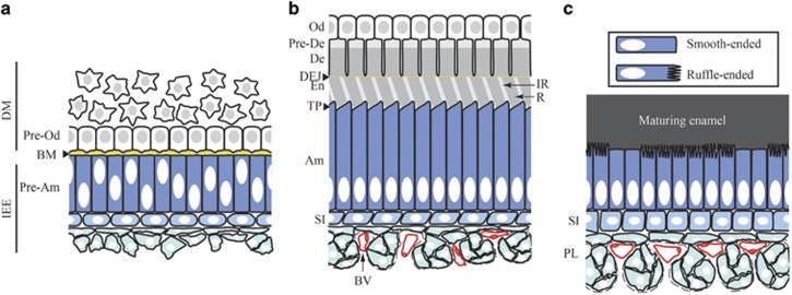
Figure 1.
Complex cellular interaction and differentiation processes involved in enamel development. The pre-secretion (a), secretion (b) and maturation (c) stages of amelogenesis are represented in the context of the tissues surrounding the developing enamel. During tooth development, the formation of enamel and dentin, the two major mineralized constituents of the tooth, is initiated at the interface between the dental mesenchyme (DM) and the inner enamel epithelium (IEE), which are separated by a basement membrane (BM). The cells from the dental mesenchyme at this interface will differentiate into odontoblasts (Od) that produce predentin (Pre-De) and drive its progression into mineralized dentin (De). In the crown, cells from the inner enamel epithelium will differentiate into enamel-producing ameloblasts (Am). Prior to enamel and dentin deposition (pre-secretion stage), interactions between pre-odontoblasts (Pre-Od) and pre-ameloblasts (Pre-Am) play a crucial role in the specification of both compartment. Pre-dentin is secreted first and is comprised mainly of type I collagen, which starts to mineralize. Pre-ameloblasts secrete enamel matrix proteins and initiate enamel mineral ribbon deposition at the dentin-enamel junction (DEJ). Ameloblasts then go through a secretory stage where they deposit enamel matrix proteins into highly structured enamel rods (R) and interrods (IR). During this stage ameloblasts are elongated and develop a specialized structure at the secretion front called the Tomes’ process (TP). The secretion phase is followed by a maturation phase during which enamel matrix proteins are degraded by proteases to leave space for the full expansion of the hydroxyapatite crystals. During this stage, ameloblasts are shorter and cycle between ruffle-ended and smooth-ended phases. The epithelial cells underlying the ameloblasts progressively develop into a stratum intermedium (SI), directly in contact with the ameloblasts, and a papillary layer (PL) populated by blood vessels (BV). Although these layers certainly play an important role in enamel development, their function remains poorly understood.

