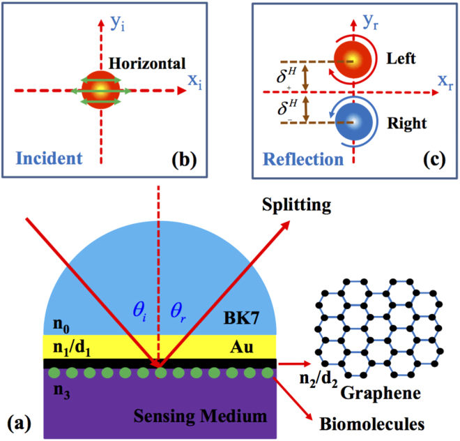
Figure 1.
Schematic of the biosensor and the corresponding photonic SHE. (a) The physical structure of the biosensor. It is composed of a BK7 glass, an Au film, and a graphene sheet. The inset shows the atomic structure of graphene. (b) The intensity and polarization distribution of the incident light beam. (c) The intensity and polarization distribution of the reflected light beam. indicate the transverse (in y direction) shifts of left- or right-circularly polarized component. To make the splitting characteristics more noticeable, we have amplified the initial spin-dependent shifts . θi and θr are the incident and reflected angle.

