Figure 4.
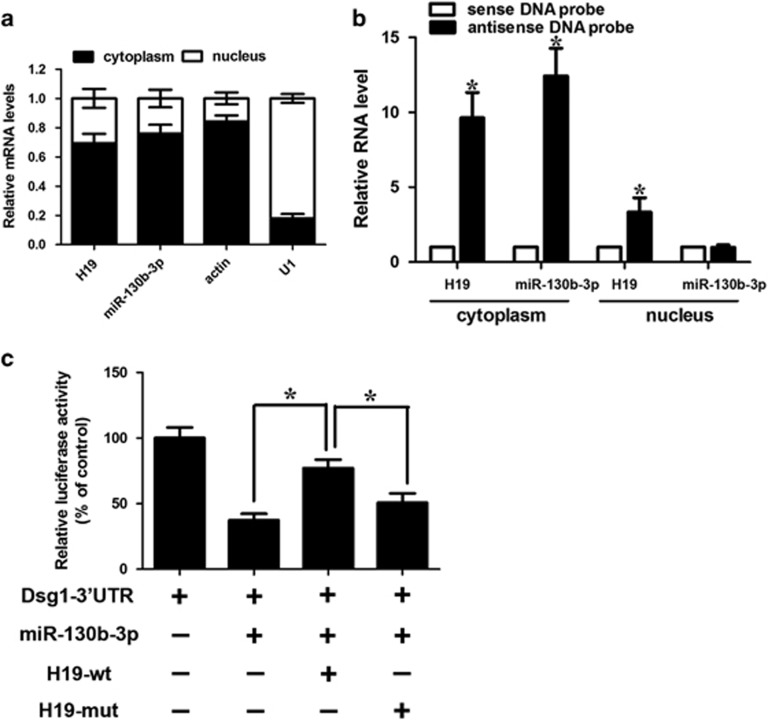
The interaction between H19 and miR-130b-3p. (a) Cellular characterization of H19, the levels of nuclear control transcript (U1), cytoplasmic control transcript (Actin) and miR-130b-3p were assessed by qRT-PCR in nuclear and cytoplasmic fractions in keratinocytes. Data are presented as a percentage of U1, Actin, H19 and miR-130b-3p levels and total levels for each were taken to be 100%. Error bars are representative of Standard deviation (S.D., n=3). (b) Keratinocytes were subjected to cytoplasm or nucleus fractionation before each fraction was incubated with in vitro-synthesized biotin-labeled sense or antisense DNA probes of H19 for biotin pull-down assay, followed by real-time RT–PCR analysis to examine miR-130b-3p levels. (c) H19 inhibits miR-130b-3p activity. Keratinocytes were infected with adenoviral H19-wt or H19-mut, then transfected with miR-130b-3p and Dsg1-3′-UTR. Luciferase activity was analyzed. Data are shown as mean±S.D. of three independent experiments. *P<0.05 in one-way analysis of variance

