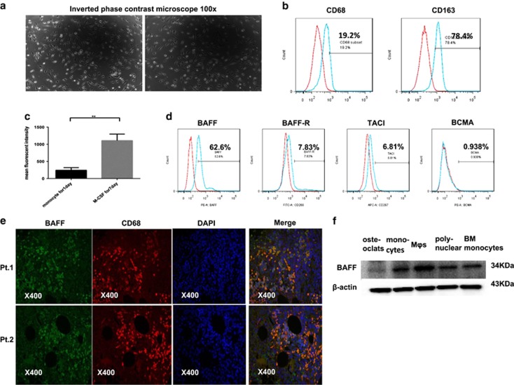
Figure 2.
PBMC-induced MΦs and the expression of BAFF and BAFF-Rs in MΦs. Monocytes from healthy donors were cultured in RPMI-1640 supplemented with 10% FBS in the presence of M-CSF (10 ng/ml) for 7 days. (a) M2-type MΦs adhered to the six-well plates and had a spindle-like morphology. (b) MΦs were positive for CD68 and CD163 by flow cytometry analysis. (c) Monocytes and monocyte-induced MΦs from seven blood donors were detected for the expression of BAFF by the flow cytometry analysis. Numbers represent the mean fluorescent intensity. (d) Cell surface expression of BAFF and its receptors in MΦs were detected by flow cytometry. (e) Expression of BAFF on primary MΦs from BM aspirates of patients with MM (n=2) were detected using immunofluorescence. (f) Expression of BAFF on monocyte-derived osteoclast, monocytes, MΦs, polynuclear cells, bone marrow monocytes were detected by Western blot

