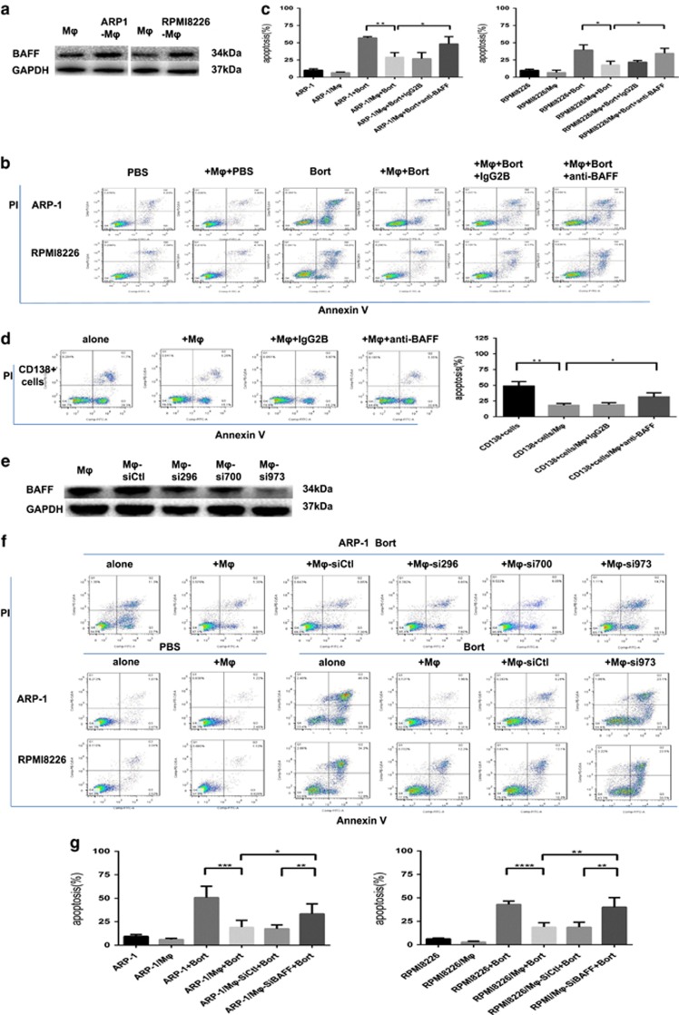
Figure 5.
BAFF is indispensable for MΦ-mediated MM bort resistance. (a) Lysates from MΦs either cultured alone or co-cultured with MM cell lines (ARP-1 and RPMI8226) were detected for the expression of BAFF protein using an anti-BAFF antibody by Western blot, with GAPDH used as a loading control. Apoptosis was evaluated by flow cytometry with Annexin V-FITC/ propidium iodide staining. (b) A representative result showing the percentage of bort-induced apoptotic MM cells (5 nM ARP-1 and 10 nM RPMI8226) in direct co-culture with MΦs, in the presence of BAFF-neutralizing antibody (20 μg/ml) or control IgG2B (20 μg/ml). (c) Reduced protective effect of MΦs in the presence of BAFF-neutralizing antibody is analyzed as means±S.D. (d) Results showing BAFF-neutralizing antibody attenuated the effect of MΦs in protecting primary CD138+ plasma cells from spontaneous apoptosis. Values are presented as means±S.D. (e) MΦs treated with BAFF-specific siRNAs showed a diverse reduction of BAFF protein compared with nontargeting siRNA (control) at 72 h using Western blot, with GAPDH as a loading control. (f) A representative result showing bort-induced apoptosis on ARP-1 cells co-cultured with MΦs following the BAFF-specific·siRNAs using flow cytometry analysis. The BAFF knockdown effect resulted in reduced ability of MΦs in protecting MM cells. (g) Result showing percentage of bort-induced apoptotic MM cells (ARP-1 and RPMI8226) in direct co-culture with BAFF-knocked down MΦs. Values are presented as means±S.D.*P<0.05, **P<0.01, ***P<0.001, ****p<0.0001

